Can DNA be useful to federally-recognized tribes?

A few federally-recognized tribes, such as the Mashantucket Pequot of Connecticut, have
considered using Native American DNA tests for enrollment purposes. For the Pequot, as for
other wealthy casino tribes, the financial stakes of enrollment are high: the Pequot disburse
monthly payments to each member totaling thousands of dollars. If DNA could exclude those
who cannot legitimately claim Pequot ancestry, the financial benefits for the remaining tribal
members would be great.
However, these Native American DNA tests rarely (if ever) identify genetic markers for
particular tribes. Because no tribe has been completely isolated from other human groups
throughout history, very few genetic markers are present only in the members of one tribe. In
all likelihood, genetic markers found in the Pequot also exist in many other tribes.
Consequently, adoption of a DNA-based enrollment policy might actually expand the number
of individuals qualifying for tribal enrollment because individuals without Pequot ancestry
could claim membership based on the shared genetic markers.
This example should serve as a red flag to tribes: enrollment policies based on DNA alone could
backfire. Furthermore, because individual identity is shaped by more than genetic ancestry,
other enrollment criteria might be better able to meet the needs of land-based tribal nations.
Reservation residence or tribal community involvement, for example, can help ensure that tribal
members are also culturally connected to the tribe and committed to its future.
Some companies may encourage the notion that genetic ancestry alone makes an Indian,
though, because there is a potentially lucrative market in such over-simplification. For
example, the DNA testing company DNAToday has teamed up with DCI America (a for-profit
tribal management consulting firm) to sell “genetic identification systems” to tribes. Their $320-
per-person photo ID cards sport computer chips and list specific DNA markers. DNAToday
advocates tribal-wide DNA testing, and claims that their product is “100% reliable in terms of
creating accurate answers” to questions of tribal enrollment.

Extracting DNA from Your Cells

Cells from the lining of your mouth come loose easily, so you will be able to collect cells containing
your DNA by swishing a liquid around in your mouth.
The cells from the lining of your mouth also come off whenever you chew food. How do you think
your body replaces the cells that come off the lining of your mouth when you eat?
To extract DNA from your cells, you will need to separate the DNA from the other types of biological
molecules in your cells. What are the other main types of large biological molecules in cells?
You will be using the same basic steps that biologists use when they extract DNA

Getting Your Sample of Cells

Obtain a cup with sports drink. You will need to get thousands of your cheek cells in the sports drink
in order to extract enough DNA to see. Therefore you should swish the sports drink around in your
mouth vigorously for at least one minute. Then spit the drink back into the cup.

Step 1: Detergent

Add a small amount of detergent to a test tube (about 0.25 mL). Put a glove on the hand you will use
to hold your test tube, not the hand you will use to pour. Now carefully pour the drink containing your
cheek cells into the test tube with detergent until the tube is half full Dishwashing detergent, like all soaps, breaks up lipids. This is why you use detergents to remove
fats (which are lipids) from dirty dishes. Adding the detergent to you cheek cell solution will break
open the cell membranes and nuclear membranes and release your DNA into the solution.

Step 2: Enzymes

Add a pinch of enzyme (meat tenderizer) to your test tube. With your gloved thumb (or palm)
covering the top of the test tube; gently invert the tube five times to mix. Let the mixture sit for at least
10 minutes. While you are waiting, you will learn about the structure of DNA. Remove your glove
and throw it in the garbage.


Why am I adding enzymes?

The nucleus of each of your cells contains multiple long strands of DNA with all the instructions to
make your entire body. If you stretched out the DNA found in one of your cells, it would be 2-3
meters long. To fit all of this DNA inside a tiny cell nucleus, the DNA is wrapped tightly around
proteins. The enzyme in meat tenderizer is a protease, which is an enzyme that cuts proteins into
small pieces. As this enzyme cuts up the proteins, the DNA will unwind and separate from the
proteins.
The protease in meat tenderizer actually comes from plants, but animals also make proteases.
Where in your body do you think you make protein-cutting enzymes?

Possible Results/Conclusions From DNA Test


When the results obtained from the standard sample from a known individual are all consistent
with or are all present in the results from the unknown crime scene sample, then the results are
considered an inclusion or nonexclusion. The term “match” is also commonly used when the test
results are consistent with the results from a known individual. That individual is included (cannot be excluded) as a possible source of the DNA found in the sample. Often, statistical frequencies regarding the rarity of the particular set of genetic information observed in the unknown
evidence sample and for a known individual are provided for various population groups.
It is possible for a falsely accused individual to be included as a source of a sample, particularly
if the test system used only tests at one or a few loci (e.g., the DQα). In this situation, additional
testing at more loci should be performed with the remaining evidence and/or DNA.
for that particular case from a legal perspective. Situations where this might apply are when the
results obtained are all consistent with the individual from whom the samples were collected


Exclusions

When the results obtained from the standard sample from a known individual are not all present in the results from the unknown crime scene sample, the results are considered an exclusion, a
nonmatch, or noninclusion. With limited exceptions, an exclusion of an individual at any one
genetic region eliminates that individual as a source of the DNA found in the sample.
In some cases where an exclusion is reported, it may be necessary to do additional testing for
that exclusion to be meaningful to the case or to provide evidence for exoneration. A situation
where this might apply is when the defendant is excluded as a donor of the DNA in a sexual
assault case, but no samples are available from the victim and/or consensual partners.


Inconclusive Result

Results may be interpreted as inconclusive for several reasons. These include situations where
no results or only partial results are obtained from the sample due to the limited amount of suitable human DNA or where results are obtained from an unknown crime scene sample but there
are no samples from known individuals available for comparison. In the latter case, the results
would be suitable for comparison once an appropriate sample for comparison is tested.


Database

RFLP-based and PCR-based databases have been constructed and are continuing to be expanded
in many laboratories throughout the United States and the world with samples from convicted
sex offenders and convicted felons, as well as samples from unsolved crimes. These databases
will be especially helpful for linking previously unrelated cases and for screening a large number
of known individuals already convicted of a crime to newly tested crime scene samples.
DNA databases of mitochondrial sequences are being established that are currently being used
for statistical purposes. It is possible that databases containing mitochondrial sequences may be
constructed for comparison to crime scene samples in the future.

Testing in the Future

Testing of hair shafts using mitochondrial DNA sequencing likely will become more widely
available in the immediate future. It may be possible to isolate and test DNA from other samples
that are not routinely tested today (e.g., fingerprints).
Y-specific probes are sequences of DNA found only on the Y (or male) chromosome. Development and validation of these probes are in progress. These probes will be especially useful for
mixed samples in which the female component is not relevant or may make interpretation of the
results more difficult (e.g., sexual assault samples, fingernails from female victims when the
assailant is male) and in the analysis and determination of the number of male sources of DNA in
samples where there are multiple male contributors (e.g., multiple assailants and/or consensual
partners in sexual assault samples). Because Y chromosomes are inherited through the male lineage, Y-specific probe results may be used to link a crime scene sample to a particular family.
DNA probes useful for identification testing are being developed from many other organisms and
may be useful in crime scene investigation. There are reported cases in which DNA from cat hair18
and from a particular type of plant has been used to link individuals to a particular crime scene.
Progress is being made in developing technologies for miniaturization of DNA tests (e.g.,
microchip analysis) that may be applied to forensic testing in the future. Expansion of existing
technologies (e.g., sequencing of nuclear DNA) may emerge for forensic testing. Other as yet
unknown or undeveloped technologies may be forthcoming that could be applied to forensic
testing. It is likely that future tests could increase the sensitivity and speed of testing, as well
as increase the discrimination capability of a test to unique identification of an individual.

Chemical structure


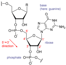
Each nucleotide in RNA contains a ribose sugar, with carbons numbered 1 through 5. A base is attached to the 1 position, generally adenine (A), cytosine (C), guanine (G) or uracil (U). Adenine and guanine are purines, cytosine and uracil are pyrimidines. A phosphate group is attached to the 3 position of one ribose and the 5 position of the next. The phosphate groups have a negative charge each at physiological pH, making RNA a charged molecule (polyanion). The bases may form hydrogen bonds between cytosine and guanine, between adenine and uracil and between guanine and uracil. However other interactions are possible, such as a group of adenine bases binding to each other in a bulge, or the GNRA tetraloop that has a guanine–adenine base-pair.

An important structural feature of RNA that distinguishes it from DNA is the presence of a hydroxyl group at the 2 position of the ribose sugar. The presence of this functional group causes the helix to adopt the A-form geometry rather than the B-form most commonly observed in DNA.This results in a very deep and narrow major groove and a shallow and wide minor groove. A second consequence of the presence of the 2-hydroxyl group is that in conformationally flexible regions of an RNA molecule (that is, not involved in formation of a double helix), it can chemically attack the adjacent phosphodiester bond to cleave the backbone

Gene Cloning and DNA Analysis

Gene cloning is the process in which a gene of interest is located and copied (cloned) out of DNA extracted from an organism,” according to the Education Center of the University of Nebraska. DNA analysis is any technique used to analyze genes and DNA. Scientists catalogue an organism’s DNA in gene libraries in order to pick up a particular genome from thousands of different genes within a DNA. DNA analysis is extensively used to determine paternity, maternity and other biological relationships among people.


HISTORY OF GENE CLONING
Hans Dreisch was the first person to create cloned animals, in the late 18th century. He wanted to prove that during cell division the genetic material is not lost. Dreisch experimented with sea urchins, which grow independently, away from their mothers, and have large embryos. A two-celled sea urchin was put into a beaker of sea water and shaken until the cells separated. Each cell grew separately and formed a sea urchin.


CLONING AT CELLULAR LEVEL
It is only recently that cloning has been done at the cellular level in the animal kingdom. In November 1951, a team of scientists working at Robert Briggs Lab cloned a frog embryo. Mammals were first cloned in 1986 by two teams working independently and using similar methods. One team, led by Neal First in the United States, cloned a cow's embryo. The other team was led by Steen Willadsen in England, and cloned a sheep’s embryo.


SHEEP CLONING
In 1986, Ian Wilmut at the Roslin Institute in Scotland was assigned a project to clone sheep. He had to create sheep that could produce certain chemicals in their milk. He altered adult cells and then cloned them. He managed to produce sheep with the altered gene throughout their bodies. The paperwork was done in 1987 and the research in 1990. Wilmut’s team learned that by starving cells, they could be forced to the GO phase, which is similar to cell hibernation. This helped to increase the survival rate of cloned cells. On July 5, 1996, a lamb cloned from a frozen mammary cell of an adult sheep was born and named Dolly.


METHODS OF CLONING
There are three main methods of cloning. One is to transfer the nucleus. In this way, an embryo is created by fusing an adult animal cell. Cloning is also done by the microinjection method. This technique makes use of the adult cell nucleus. The third method of cloning is artificial twinning, which is a method in which the embryo is split into two or more embryos.


DNA ANALYSIS
Alec Jeffery, an English geneticist, was among the scientists who discovered DNA analysis technique. All living things have unique genetic codes. By analyzing its genetic sequences, you can identify any type of organism. DNA analysis helps scientists record an individual specimen's personality. With its use, scientists can conduct research on the evolution of a species and provide insight into our future health. DNA analysis has helped immensely in forensic science.

Transgenic organisms

Another important controversy is the possibility of unforeseen local and global effects as a result of transgenic organisms proliferating. The basic ethical issues involved in genetic research are discussed in the article on genetic engineering.Some critics have raised the concern that conventionally-bred crop plants can be cross-pollinated (bred) from the pollen of modified plants. Pollen can be dispersed over large areas by wind, animals, and insects. In 2007, the U.S. Department of Agriculture fined Scotts Miracle-Gro $500,000 when modified genetic material from creeping bentgrass, a new golf-course grass Scotts had been testing, was found within close relatives of the same genus (Agrostis) as well as in native grasses up to 21 km (13 miles) away from the test sites, released when freshly cut grass was blown by the wind.[59]GM proponents point out that outcrossing, as this process is known, is not new. The same thing happens with any new open-pollinated crop variety—newly introduced traits can potentially cross out into neighboring crop plants of the same species and, in some cases, to closely related wild relatives. Defenders of GM technology point out that each GM crop is assessed on a case-by-case basis to determine if there is any risk associated with the outcrossing of the GM trait into wild plant populations. The fact that a GM plant may outcross with a related wild relative is not, in itself, a risk unless such an occurrence has negative consequences. If, for example, an herbicide resistance trait was to cross into a wild relative of a crop plant it can be predicted that this would not have any consequences except in areas where herbicides are sprayed, such as a farm. In such a setting the farmer can manage this risk by rotating herbicides.The European Union funds research programs such as Co-Extra, that investigate options and technologies on the coexistence of GM and conventional farming. This also includes research on biological containment strategies and other measures to prevent outcrossing and enable the implementation of coexistence.If patented genes are outcrossed, even accidentally, to other commercial fields and a person deliberately selects the outcrossed plants for subsequent planting then the patent holder has the right to control the use of those crops. This was supported in Canadian law in the case of Monsanto Canada Inc. v. Schmeiser.

DNA TECHNOLOGY FOR FORENSIC INVESTIGATION

RFLP was one of the aboriginal applications of DNA assay to argumentative investigation. With the development of newer, added able DNA-analysis techniques, RFLP is not acclimated as abundant as it already was because it requires almost ample amounts of DNA. In addition, samples base by ecology factors, such as clay or mold, do not assignment able-bodied with RFLP.PCR AnalysisPolymerase alternation acknowledgment (PCR) is acclimated to accomplish millions of exact copies of DNA from a biological sample. DNA addition with PCR allows DNA assay on biological samples as baby as a few bark cells. With RFLP, DNA samples would accept to be about the admeasurement of a quarter. The adeptness of PCR to amplify such tiny quantities of DNA enables alike awful base samples to be analyzed. Great care, however, charge be taken to anticipate contagion with added biological abstracts during the identifying, collecting, and attention of a sample.STR AnalysisShort bike echo (STR) technology is acclimated to appraise specific regions (loci) aural nuclear DNA. Variability in STR regions can be acclimated to analyze one DNA contour from another. The Federal Bureau of Assay (FBI) uses a accepted set of 13 specific STR regions for CODIS. CODIS is a software affairs that operates local, state, and civic databases of DNA profiles from bedevilled offenders, baffling abomination arena evidence, and missing persons. The allowance that two individuals will accept the aforementioned 13-loci DNA contour is about one in a billion.Mitochondrial DNA AnalysisMitochondrial DNA assay (mtDNA) can be acclimated to appraise the DNA from samples that cannot be analyzed by RFLP or STR. Nuclear DNA charge be extracted from samples for use in RFLP, PCR, and STR; however, mtDNA assay uses DNA extracted from addition cellular organelle alleged a mitochondrion. While earlier biological samples that abridgement nucleated cellular material, such as hair, bones, and teeth, cannot be analyzed with STR and RFLP, they can be analyzed with mtDNA. In the assay of cases that accept gone baffling for abounding years, mtDNA is acutely valuable.All mothers accept the aforementioned mitochondrial DNA as their offspring. This is because the mitochondria of anniversary new antecedent comes from the mother's egg cell. The father's agent contributes alone nuclear DNA. Comparing the mtDNA contour of anonymous charcoal with the contour of a abeyant affectionate about can be an important address in missing-person investigations.Y-Chromosome AnalysisThe Y chromosome is anesthetized anon from ancestor to son, so assay of abiogenetic markers on the Y chromosome is abnormally advantageous for archetype relationships amid males or for allegory biological affirmation involving assorted macho contributors.

fingerprinting

DNA fingerprinting, also known as DNA typing, is a method of isolating and making images of sequences of DNA. The technique was developed in 1984 by the British geneticist Alec Jeffreys, after he noticed the existence of certain sequences of DNA (called minisatellites) that do not contribute to the function of a gene but are repeated within the gene and in other genes of a DNA sample. Jeffreys also determined that each organism has a unique pattern of these minisatellites, the only exception being multiple individuals from a single zygote (e.g., identical twins). The procedure for creating a DNA fingerprint consists of first obtaining a sample of cells containing DNA (e.g., from skin, blood, or hair), extracting the DNA, and purifying it. The DNA is then cut at specific points along the strand with substances called restriction enzymes. This produces fragments of varying lengths that are sorted by placing them on a gel and then subjecting the gel toan electric current (electrophoresis): the shorter the fragment the more quickly it will move toward the positive pole (anode). The sorted, double-stranded DNA fragments are then subjected to a blotting technique in which they are split into single strands and transferred to a nylon sheet. The fragments undergo autoradiography in which they are exposed to DNA probes—pieces of synthetic DNA that have been made radioactive and that bind to the minisatellites. A piece of X-ray film is then exposed to the fragments, and a dark mark is produced at any point where a radioactive probe has become attached. The resultant pattern of these marks can then be analyzed. An early use of DNA fingerprinting was in legal disputes,notably to help solve crimes and to determine paternity. The technique was challenged, however, overconcerns about sample contamination, faulty preparation procedures, and erroneous interpretation of the results. Efforts were made to improve reliability, and today the technique has been refined through the use of more specific and more sensitive probes and better blotting membranes. It also has been recognized that DNA fingerprinting, similar to other DNA analysis techniques, is limited by the quality of the sample obtained. DNA samples that are degraded or collected postmortem typicallyproduce less reliable results than do samples that are obtained from a living individual. If only a small amount of DNA is available for fingerprinting, PCR may be used to create thousands of copies of a DNA segment. Once an adequate amount of DNA has been produced, the exact sequence of nucleotide pairs in a segment of DNA can be determined using one of several biomolecular sequencing methods. Automated equipment has greatly increased the speed of DNA sequencing and has made available many practical applications, including pinpointing segments of genes that cause genetic diseases, mapping the human genome, engineering drought-resistant plants, and producing biological drugs from genetically altered bacteria.

human genetics

Most of the emphasis is on understanding and treating genetic disease and genetically influenced ill health, areas collectively known as medical genetics. One broad area of activity is laboratory research dealing with the mechanisms of human gene function and malfunction and investigating pharmaceutical and other types of treatments. Since there is a high degree of evolutionary conservation between organisms, research on model organisms—such as bacteria, fungi, and fruit flies (Drosophila)—which are easier to study, often provides important insights into human gene function. Many single-gene diseases, caused by mutant alleles of a single gene, have been discovered. Two well-characterized single-gene diseases include phenylketonuria (PKU) and Tay-Sachs disease. Other diseases, such as heart disease, schizophrenia, and depression, are thought to have morecomplex heredity components that involve a number of different genes. These diseases are the focus of a great deal of research that is being carried out today. In addition,abnormalities in chromosomes have been identified by studies employing techniques such as chromosomal banding. Individual chromosomes are identified by the banding patterns revealed by different staining techniques.Segments of chromosomes or chromosomes that are aberrant in number and morphology may be precisely identified.Another broad area of activity is clinical genetics, which centres on advising parents of the likelihood of their children being affected by genetic disease caused by mutant genes and abnormal chromosome structure and umber. Such genetic counseling is based on examining individual and family medical records and on diagnosticprocedures that can detect unexpressed, abnormal forms of genes. Counseling is carried out by physicians with a particular interest in this area or by specially trained nonphysicians.

RNA data sequences to DNA

Proteins are not the only substances that are synthesized directly from data within the DNA. Some forms of RNA are specialized, and also have their formula encoded directly in digital DNA formulae. Not all types of RNA are temporary intermediate forms with their form depending on whatever DNA they are copying. There are certain forms of RNA that have a particular form that is the same across all individuals. Some of these special-purpose RNA forms are,
1. tRNA - transfer RNA
2. rRNA - ribosome RNA

There are exactly 20 forms of tRNA, one each transfer a particular amino acid. tRNA molecules contain about 75-80 bases. tRNA recognizes one of the 64 triplets, and matches it one of the 20 amino acids. Since there are 20 tRNA types, and not 64, each tRNA molecule has to recognize more than one triplet ordering as a match. The DNA code contains multiple repetitions of codes for tRNA and rRNA. About 280 copies are spread over 5 chromosomes. Presumably, this allows each cell to make multiple copies of tRNA and rRNA molecules at once from its single copy of the DNA.

RECOMBINANT DNA

Recombinant DNA is a form of artificial DNA that is engineered through the combination of insertion of one or more DNA strands. Therefor combining DNA sequences that would not normally occur together. In terms of genetic modification, recombinant DNA is produced through the addition of relevant DNA into an existing organismal genome, such as the plasmid of bacteria, to code for or alter different traits for a specific purpose, such as immunity. It differs from genetic recombination, in that it does not occur through processes within the cell or ribosome, but is exclusively engineered.The Recombinant DNA technique was engineered by Stanley Norman Cohen and Herbert Boyer in 1973. They published their findings in a 1974 paper entitled "Construction of Biologically Functional Bacterial Plasmids in vitro". Which described a technique to isolate and amplify genes or DNA segments and insert them into another cell with precision, creating a transgenic bacterium.

Phylogetics DNA

Every living cells contains DNA, RNA and protiens. closely related organisms generally have degree of agreement in the molecular structure of these substances,Heterogenous nuclear ribonucleoprotiens are spliceosomal macromolecular assemblages and thus actively participate in pre-mRNA metabloism.

conserved sequnces. such as mitochondrial DNA are expected to accumalate mutations over time and assuming a constant rate of mutation provide molecular interaction features.

Molecular characterization of human hnRNP A3 showed that while the recombinant hnRNP A3 with its 296 amino acids migrates as excpted as 32 kDA protien on SDS-PAGE analysis, it is recoginzed by the patient's sera as a 50 kda highly related ye unknownv crossreactive protien.

The sequence comparision. however may be insufficient for deduction of its functional role patients diagnozed with the suspected disease. therefore we followed the suggestion of ponting CS and Russel RRSurprisingly, neither the 50 kda nor the 32kda protien was dected in the suspected disease.

GENETIC DISORDER

Genetic disorder is a condition caused by abnormalities in genes or chromosomes. While some diseases such as cancer. Cancer are due to genetic abnormalities acquired in a few cells during life. The term "genetic disease" most commonly refers to diseases present in all cells of the body and present since conception. Some genetic disorders are caused by chromosomal abnormalities due to errors in meiosis, the process which produces reproductive cells such as sperm and eggs. Examples include Down syndrome, Turner Syndrome and Klinefelter's syndrome. Other genetic changes may occur during the production of germ cells by the parent. One example is the triplet expansion repeat mutations which can cause fragile X syndrome or Huntington's disease. Defective genes may also be inherited intact from the parents. In this case, the genetic disorder is known as a hereditary disease. This can often happen unexpectedly when two healthy carriers of a defective recessive gene reproduce, but can also happen when the defective gene is dominant.

About 4,000 genetic disorders are known with more being discovered. Most disorders are quite rare and affect one person in every several thousands or millions. Cystic fibrosis is one of the most common genetic disorders around 5% of the population of the United States carry at least one copy of the defective gene. Some types of recessive gene disorder confer an advantage in the heterozygous state in certain environments.Genetic diseases are typically diagnosed and treated by geneticists. Genetic counselors assist the physicians and directly counsel patients. The study of genetic diseases is a scientific discipline whose theoretical underpinning is based on population genetics.

DNA (DEOXYRIBOSE NUCLEIC ACID)

DNA strand is made from alternating phospate and sugar residues. Thesre two strands run in opposite directions to each other and therfore antiparallel Attached to each sugar is one of four types of molecules called bases. The sugar in DNA is 2 - deoxyribose which is apentose sugar searches of DNA into the related nuclear acid RNA in aproesss sugsrs are joinrd to phospate groups that from phosphodiester bonds btwn the third and4th carbon atoms adjacent sugar rings.

DNA is a long polymer made from repeating units called nucleotides. The DNA is chain is 20 - 26 angstroms wide. DNA polymers can be very large moleculars containing millions of nucleotides chemically with backbones made of sugars and phospates groups joined by ester bonds these onformation is using the genetic code. Phospate groups are joined by ester bonds. In living organisms.

DNA does not usually exist as a single molecule but it exists as a pair of molecules that are held tightly together. These two long strands and entwine like vines in the shape a double helix. which holds chain together and a base which interacts with the other DNA strand in the helix. A base linked to a sugar is nucleiotide. If muultiple nucleiotide are linked to together as in DNA this polymer is known as a polynucleiotide

History of DNA research

DNA was first isolated by the Swiss physician Friedrich Miescher who, in 1869, discovered a microscopic substance in the pus of discarded surgical bandages. As it resided in the nuclei of cells, he called it "nuclein". In 1919, Phoebus Levene identified the base, sugar and phosphate nucleotide unit. Levene suggested that DNA consisted of a string of nucleotide units linked together through the phosphate groups. However, Levene thought the chain was short and the bases repeated in a fixed order. In 1937 William Astbury produced the first X-ray diffraction patterns that showed that DNA had a regular structure.

In 1928, Frederick Griffith discovered that traits of the "smooth" form of the Pneumococcus could be transferred to the "rough" form of the same bacteria by mixing killed "smooth" bacteria with the live "rough" form. This system provided the first clear suggestion that DNA carried genetic information—the Avery-MacLeod-McCarty experiment—when Oswald Avery, along with coworkers Colin MacLeod and Maclyn McCarty, identified DNA as the transforming principle in 1943. DNA's role in heredity was confirmed in 1952, when Alfred Hershey and Martha Chase in the Hershey-Chase experiment showed that DNA is the genetic material of the T2 phage.


Francis Crick


Rosalind Franklin


Raymond Gosling


In 1953 James D. Watson and Francis Crick suggested what is now accepted as the first correct double-helix model of DNA structure in the journal Nature. Their double-helix, molecular model of DNA was then based on a single X-ray diffraction image taken by Rosalind Franklin and Raymond Gosling in May 1952, as well as the information that the DNA bases were paired—also obtained through private communications from Erwin Chargaff in the previous years. Chargaff's rules played a very important role in establishing double-helix configurations for B-DNA as well as A-DNA.

Experimental evidence supporting the Watson and Crick model were published in a series of five articles in the same issue of Nature. Of these, Franklin and Gosling's paper was the first publication of their own X-ray diffraction data and original analysis method that partially supported the Watson and Crick mode; this issue also contained an article on DNA structure by Maurice Wilkins and two of his colleagues, whose analysis and in vivo B-DNA X-ray patterns also supported the presence in vivo of the double-helical DNA configurations as proposed by Crick and Watson for their double-helix molecular model of DNA in the previous two pages of Nature. In 1962, after Franklin's death, Watson, Crick, and Wilkins jointly received the Nobel Prize in Physiology or Medicine. Unfortunately, Nobel rules of the time allowed only living recipients, but a vigorous debate continues on who should receive credit for the discovery.
In an influential presentation in 1957, Crick laid out the "Central Dogma" of molecular biology, which foretold the relationship between DNA, RNA, and proteins, and articulated the "adaptor hypothesis". Final confirmation of the replication mechanism that was implied by the double-helical structure followed in 1958 through the Meselson-Stahl experiment. Further work by Crick and coworkers showed that the genetic code was based on non-overlapping triplets of bases, called codons, allowing Har Gobind Khorana, Robert W. Holley and Marshall Warren Nirenberg to decipher the genetic code. These findings represent the birth of molecular biology

DNA nanotechnology

DNA nanotechnology uses the unique molecular recognition properties of DNA and other nucleic acids to create self-assembling branched DNA complexes with useful properties.[124] DNA is thus used as a structural material rather than as a carrier of biological information. This has led to the creation of two-dimensional periodic lattices (both tile-based as well as using the "DNA origami" method) as well as three-dimensional structures in the shapes of polyhedra. Nanomechanical devices and algorithmic self-assembly have also been demonstrated, and these DNA structures have been used to template the arrangement of other molecules such as gold nanoparticles and streptavidin proteins.

Bioinformatics

Bioinformatics involves the manipulation, searching, and data mining of DNA sequence data. The development of techniques to store and search DNA sequences have led to widely applied advances in computer science, especially string searching algorithms, machine learning and database theory.[120] String searching or matching algorithms, which find an occurrence of a sequence of letters inside a larger sequence of letters, were developed to search for specific sequences of nucleotides. In other applications such as text editors, even simple algorithms for this problem usually suffice, but DNA sequences cause these algorithms to exhibit near-worst-case behaviour due to their small number of distinct characters. The related problem of sequence alignment aims to identify homologous sequences and locate the specific mutations that make them distinct. These techniques, especially multiple sequence alignment, are used in studying phylogenetic relationships and protein function. Data sets representing entire genomes' worth of DNA sequences, such as those produced by the Human Genome Project, are difficult to use without annotations, which label the locations of genes and regulatory elements on each chromosome. Regions of DNA sequence that have the characteristic patterns associated with protein- or RNA-coding genes can be identified by gene finding algorithms, which allow researchers to predict the presence of particular gene products in an organism even before they have been isolated experimentally.

Forensics

Forensic scientists can use DNA in blood, semen, skin, saliva or hair found at a crime scene to identify a matching DNA of an individual, such as a perpetrator. This process is called genetic fingerprinting, or more accurately, DNA profiling. In DNA profiling, the lengths of variable sections of repetitive DNA, such as short tandem repeats and minisatellites, are compared between people. This method is usually an extremely reliable technique for identifying a matching DNA. However, identification can be complicated if the scene is contaminated with DNA from several people. DNA profiling was developed in 1984 by British geneticist Sir Alec Jeffreys, and first used in forensic science to convict Colin Pitchfork in the 1988 Enderby murders case.

People convicted of certain types of crimes may be required to provide a sample of DNA for a database. This has helped investigators solve old cases where only a DNA sample was obtained from the scene. DNA profiling can also be used to identify victims of mass casualty incidents. On the other hand, many convicted people have been released from prison on the basis of DNA techniques, which were not available when a crime had originally been committed.

Genetic engineering

Methods have been developed to purify DNA from organisms, such as phenol-chloroform extraction and manipulate it in the laboratory, such as restriction digests and the polymerase chain reaction. Modern biology and biochemistry make intensive use of these techniques in recombinant DNA technology. Recombinant DNA is a man-made DNA sequence that has been assembled from other DNA sequences. They can be transformed into organisms in the form of plasmids or in the appropriate format, by using a viral vector. The genetically modified organisms produced can be used to produce products such as recombinant proteins, used in medical research, or be grown in agriculture.

DNA-binding proteins

Structural proteins that bind DNA are well-understood examples of non-specific DNA-protein interactions. Within chromosomes, DNA is held in complexes with structural proteins. These proteins organize the DNA into a compact structure called chromatin. In eukaryotes this structure involves DNA binding to a complex of small basic proteins called histones, while in prokaryotes multiple types of proteins are involved. The histones form a disk-shaped complex called a nucleosome, which contains two complete turns of double-stranded DNA wrapped around its surface. These non-specific interactions are formed through basic residues in the histones making ionic bonds to the acidic sugar-phosphate backbone of the DNA, and are therefore largely independent of the base sequence. Chemical modifications of these basic amino acid residues include methylation, phosphorylation and acetylation. These chemical changes alter the strength of the interaction between the DNA and the histones, making the DNA more or less accessible to transcription factors and changing the rate of transcription. Other non-specific DNA-binding proteins in chromatin include the high-mobility group proteins, which bind to bent or distorted DNA. These proteins are important in bending arrays of nucleosomes and arranging them into the larger structures that make up chromosomes.

A distinct group of DNA-binding proteins are the DNA-binding proteins that specifically bind single-stranded DNA. In humans, replication protein A is the best-understood member of this family and is used in processes where the double helix is separated, including DNA replication, recombination and DNA repair. These binding proteins seem to stabilize single-stranded DNA and protect it from forming stem-loops or being degraded by nucleases.
The lambda repressor helix-turn-helix transcription factor bound to its DNA target

In contrast, other proteins have evolved to bind to particular DNA sequences. The most intensively studied of these are the various transcription factors, which are proteins that regulate transcription. Each transcription factor binds to one particular set of DNA sequences and activates or inhibits the transcription of genes that have these sequences close to their promoters. The transcription factors do this in two ways. Firstly, they can bind the RNA polymerase responsible for transcription, either directly or through other mediator proteins; this locates the polymerase at the promoter and allows it to begin transcription. Alternatively, transcription factors can bind enzymes that modify the histones at the promoter; this will change the accessibility of the DNA template to the polymerase.

As these DNA targets can occur throughout an organism's genome, changes in the activity of one type of transcription factor can affect thousands of genes. Consequently, these proteins are often the targets of the signal transduction processes that control responses to environmental changes or cellular differentiation and development. The specificity of these transcription factors' interactions with DNA come from the proteins making multiple contacts to the edges of the DNA bases, allowing them to "read" the DNA sequence. Most of these base-interactions are made in the major groove, where the bases are most accessible.

Transcription and translation

A gene is a sequence of DNA that contains genetic information and can influence the phenotype of an organism. Within a gene, the sequence of bases along a DNA strand defines a messenger RNA sequence, which then defines one or more protein sequences. The relationship between the nucleotide sequences of genes and the amino-acid sequences of proteins is determined by the rules of translation, known collectively as the genetic code. The genetic code consists of three-letter 'words' called codons formed from a sequence of three nucleotides (e.g. ACT, CAG, TTT).

In transcription, the codons of a gene are copied into messenger RNA by RNA polymerase. This RNA copy is then decoded by a ribosome that reads the RNA sequence by base-pairing the messenger RNA to transfer RNA, which carries amino acids. Since there are 4 bases in 3-letter combinations, there are 64 possible codons (43 combinations). These encode the twenty standard amino acids, giving most amino acids more than one possible codon. There are also three 'stop' or 'nonsense' codons signifying the end of the coding region; these are the TAA, TGA and TAG codons.

Genes and genomes

Genomic DNA is located in the cell nucleus of eukaryotes, as well as small amounts in mitochondria and chloroplasts. In prokaryotes, the DNA is held within an irregularly shaped body in the cytoplasm called the nucleoid. The genetic information in a genome is held within genes, and the complete set of this information in an organism is called its genotype. A gene is a unit of heredity and is a region of DNA that influences a particular characteristic in an organism. Genes contain an open reading frame that can be transcribed, as well as regulatory sequences such as promoters and enhancers, which control the transcription of the open reading frame.

In many species, only a small fraction of the total sequence of the genome encodes protein. For example, only about 1.5% of the human genome consists of protein-coding exons, with over 50% of human DNA consisting of non-coding repetitive sequences. The reasons for the presence of so much non-coding DNA in eukaryotic genomes and the extraordinary differences in genome size, or C-value, among species represent a long-standing puzzle known as the "C-value enigma." However, DNA sequences that do not code protein may still encode functional non-coding RNA molecules, which are involved in the regulation of gene expression.
T7 RNA polymerase (blue) producing a mRNA (green) from a DNA template (orange).

Some non-coding DNA sequences play structural roles in chromosomes. Telomeres and centromeres typically contain few genes, but are important for the function and stability of chromosomes. An abundant form of non-coding DNA in humans are pseudogenes, which are copies of genes that have been disabled by mutation. These sequences are usually just molecular fossils, although they can occasionally serve as raw genetic material for the creation of new genes through the process of gene duplication and divergence.

Biological functions

DNA usually occurs as linear chromosomes in eukaryotes, and circular chromosomes in prokaryotes. The set of chromosomes in a cell makes up its genome; the human genome has approximately 3 billion base pairs of DNA arranged into 46 chromosomes.[64] The information carried by DNA is held in the sequence of pieces of DNA called genes. Transmission of genetic information in genes is achieved via complementary base pairing. For example, in transcription, when a cell uses the information in a gene, the DNA sequence is copied into a complementary RNA sequence through the attraction between the DNA and the correct RNA nucleotides. Usually, this RNA copy is then used to make a matching protein sequence in a process called translation which depends on the same interaction between RNA nucleotides. Alternatively, a cell may simply copy its genetic information in a process called DNA replication. The details of these functions are covered in other articles; here we focus on the interactions between DNA and other molecules that mediate the function of the genome.

Quadruplex structures

At the ends of the linear chromosomes are specialized regions of DNA called telomeres. The main function of these regions is to allow the cell to replicate chromosome ends using the enzyme telomerase, as the enzymes that normally replicate DNA cannot copy the extreme 3′ ends of chromosomes. These specialized chromosome caps also help protect the DNA ends, and stop the DNA repair systems in the cell from treating them as damage to be corrected. In human cells, telomeres are usually lengths of single-stranded DNA containing several thousand repeats of a simple TTAGGG sequence.

These guanine-rich sequences may stabilize chromosome ends by forming structures of stacked sets of four-base units, rather than the usual base pairs found in other DNA molecules. Here, four guanine bases form a flat plate and these flat four-base units then stack on top of each other, to form a stable G-quadruplex structure. These structures are stabilized by hydrogen bonding between the edges of the bases and chelation of a metal ion in the centre of each four-base unit. Other structures can also be formed, with the central set of four bases coming from either a single strand folded around the bases, or several different parallel strands, each contributing one base to the central structure.

In addition to these stacked structures, telomeres also form large loop structures called telomere loops, or T-loops. Here, the single-stranded DNA curls around in a long circle stabilized by telomere-binding proteins. At the very end of the T-loop, the single-stranded telomere DNA is held onto a region of double-stranded DNA by the telomere strand disrupting the double-helical DNA and base pairing to one of the two strands. This triple-stranded structure is called a displacement loop

Alternate DNA structures

DNA exists in many possible conformations that include A-DNA, B-DNA, and Z-DNA forms, although, only B-DNA and Z-DNA have been directly observed in functional organisms. The conformation that DNA adopts depends on the hydration level, DNA sequence, the amount and direction of supercoiling, chemical modifications of the bases, the type and concentration of metal ions, as well as the presence of polyamines in solution.

The first published reports of A-DNA X-ray diffraction patterns— and also B-DNA used analyses based on Patterson transforms that provided only a limited amount of structural information for oriented fibers of DNA. An alternate analysis was then proposed by Wilkins et al., in 1953, for the in vivo B-DNA X-ray diffraction/scattering patterns of highly hydrated DNA fibers in terms of squares of Bessel functions. In the same journal, Watson and Crick presented their molecular modeling analysis of the DNA X-ray diffraction patterns to suggest that the structure was a double-helix.

Although the `B-DNA form' is most common under the conditions found in cells, it is not a well-defined conformation but a family of related DNA conformations that occur at the high hydration levels present in living cells. Their corresponding X-ray diffraction and scattering patterns are characteristic of molecular paracrystals with a significant degree of disorder.

Compared to B-DNA, the A-DNA form is a wider right-handed spiral, with a shallow, wide minor groove and a narrower, deeper major groove. The A form occurs under non-physiological conditions in partially dehydrated samples of DNA, while in the cell it may be produced in hybrid pairings of DNA and RNA strands, as well as in enzyme-DNA complexes. Segments of DNA where the bases have been chemically modified by methylation may undergo a larger change in conformation and adopt the Z form. Here, the strands turn about the helical axis in a left-handed spiral, the opposite of the more common B form. These unusual structures can be recognized by specific Z-DNA binding proteins and may be involved in the regulation of transcription

Sense and antisense

A DNA sequence is called "sense" if its sequence is the same as that of a messenger RNA copy that is translated into protein. The sequence on the opposite strand is called the "antisense" sequence. Both sense and antisense sequences can exist on different parts of the same strand of DNA (i.e. both strands contain both sense and antisense sequences). In both prokaryotes and eukaryotes, antisense RNA sequences are produced, but the functions of these RNAs are not entirely clear. One proposal is that antisense RNAs are involved in regulating gene expression through RNA-RNA base pairing.

A few DNA sequences in prokaryotes and eukaryotes, and more in plasmids and viruses, blur the distinction between sense and antisense strands by having overlapping genes. In these cases, some DNA sequences do double duty, encoding one protein when read along one strand, and a second protein when read in the opposite direction along the other strand. In bacteria, this overlap may be involved in the regulation of gene transcription, while in viruses, overlapping genes increase the amount of information that can be encoded within the small viral genome.

Base pairing

Each type of base on one strand forms a bond with just one type of base on the other strand. This is called complementary base pairing. Here, purines form hydrogen bonds to pyrimidines, with A bonding only to T, and C bonding only to G. This arrangement of two nucleotides binding together across the double helix is called a base pair. As hydrogen bonds are not covalent, they can be broken and rejoined relatively easily. The two strands of DNA in a double helix can therefore be pulled apart like a zipper, either by a mechanical force or high temperature. As a result of this complementarity, all the information in the double-stranded sequence of a DNA helix is duplicated on each strand, which is vital in DNA replication. Indeed, this reversible and specific interaction between complementary base pairs is critical for all the functions of DNA in living organisms.
GC DNA base pair.svg
AT DNA base pair.svg
Top, a GC base pair with three hydrogen bonds. Bottom, an AT base pair with two hydrogen bonds. Non-covalent hydrogen bonds between the pairs are shown as dashed lines.

The two types of base pairs form different numbers of hydrogen bonds, AT forming two hydrogen bonds, and GC forming three hydrogen bonds (see figures, left). DNA with high GC-content is more stable than DNA with low GC-content, but contrary to popular belief, this is not due to the extra hydrogen bond of a GC base pair but rather the contribution of stacking interactions (hydrogen bonding merely provides specificity of the pairing, not stability). As a result, it is both the percentage of GC base pairs and the overall length of a DNA double helix that determine the strength of the association between the two strands of DNA. Long DNA helices with a high GC content have stronger-interacting strands, while short helices with high AT content have weaker-interacting strands. In biology, parts of the DNA double helix that need to separate easily, such as the TATAAT Pribnow box in some promoters, tend to have a high AT content, making the strands easier to pull apart. In the laboratory, the strength of this interaction can be measured by finding the temperature required to break the hydrogen bonds, their melting temperature (also called Tm value). When all the base pairs in a DNA double helix melt, the strands separate and exist in solution as two entirely independent molecules. These single-stranded DNA molecules have no single common shape, but some conformations are more stable than others

Grooves

Twin helical strands form the DNA backbone. Another double helix may be found by tracing the spaces, or grooves, between the strands. These voids are adjacent to the base pairs and may provide a binding site. As the strands are not directly opposite each other, the grooves are unequally sized. One groove, the major groove, is 22 Å wide and the other, the minor groove, is 12 Å wide. The narrowness of the minor groove means that the edges of the bases are more accessible in the major groove. As a result, proteins like transcription factors that can bind to specific sequences in double-stranded DNA usually make contacts to the sides of the bases exposed in the major groove. This situation varies in unusual conformations of DNA within the cell (see below), but the major and minor grooves are always named to reflect the differences in size that would be seen if the DNA is twisted back into the ordinary B form